Case of Spontaneous Hemoperitoneum in Chronic Liver Disease
A 53-year-old male with a known history of chronic liver disease (CLD) presented with low hemoglobin levels but without any history of hematemesis or melena. This situation was concerning for physicians as it necessitated the identification of a potential source of bleeding. The patient required four units of packed cell volume (PCV) and eight units of fresh frozen plasma (FFP) for stabilization.
A few months later, the patient experienced a recurrence of similar symptoms, necessitating hospitalization and blood transfusions once again. Despite these interventions, the cause of bleeding remained unidentified, leading to a referral to me, Dr. Keyur Bhatt. A CT scan had previously been performed, but it did not clearly indicate the source of bleeding. Although the radiologist noted the presence of hemoperitoneum, the cause remained uncertain.
Given the clinical scenario and the lack of an identifiable source of bleeding, I recommended proceeding with diagnostic laparoscopy for further evaluation and management.
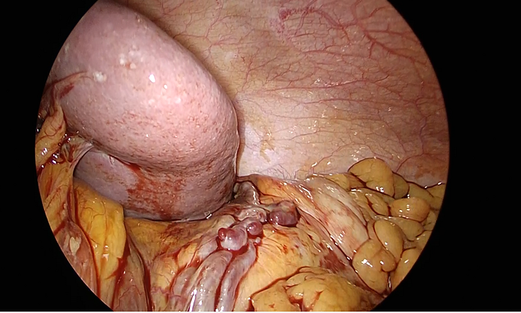
During the laparoscopic procedure, I encountered findings that were consistent with the CT scan results, which was a gratifying confirmation of my diagnostic skills. The laparoscopic exploration revealed approximately 2.2 liters of hemoperitoneum. There were no visible collaterals throughout the abdomen, and no significant retroperitoneal microcollaterals were observed in the right paracolic gutter or the pelvis.
On the left side, however, multiple collaterals, numbering between 2 and 3, were identified, extending from the splenic vein into the retroperitoneum. These collaterals exhibited varices and active bleeding. The bleeding was effectively controlled using a Ligasure device.
This case is noteworthy due to the rarity of such occurrences, highlighting the complexities and potential complications associated with chronic liver disease. The use of advanced surgical techniques was crucial in managing this emergency and ensuring a successful outcome.
This case underscores the unpredictable nature of chronic liver disease and the need for continued research and awareness to better understand and manage its complications. The local medical community is encouraged to share knowledge and experiences to improve patient outcomes in similar scenarios.
Additionally, this case emphasizes the importance for surgeons to be proficient in interpreting CT scans and integrating that knowledge into their clinical practice.