Discovering Gallbladder Lipoma: A Rare Case Study
When it comes to diagnosing and treating gallbladder abnormalities, most practitioners are familiar with conditions such as gallstones, cholecystitis, or gallbladder cancer. However, there are rarer conditions that can pose diagnostic challenges, such as gallbladder lipoma. In this blog post, we’ll delve into a fascinating case study of a gallbladder lipoma, shedding light on this rare finding and its implications for clinical practice.
What is a Gallbladder Lipoma?
A gallbladder lipoma is a benign tumor composed of adipose (fat) tissue. These tumors are quite rare, especially when compared to more common gallbladder pathologies. Lipomas are generally slow-growing and often asymptomatic, making them a challenging diagnosis during routine evaluations or imaging.
Case Study Overview:
In this particular case, the patient presented with nonspecific symptoms, which could have been attributed to a range of gallbladder disorders. The diagnosis was made incidentally during imaging studies performed for other reasons. Here’s a closer look at the key aspects of this case
1)Patient Presentation: The patient initially sought medical advice due to vague abdominal discomfort. Symptoms were not specific to gallbladder pathology, making it difficult to pinpoint the issue.
2)
Diagnostic Imaging: An abdominal ultrasound, typically the first imaging modality for evaluating gallbladder conditions, was performed. While ultrasound is excellent for detecting gallstones and inflammation, it sometimes struggles with differentiating certain types of tumors.
3)
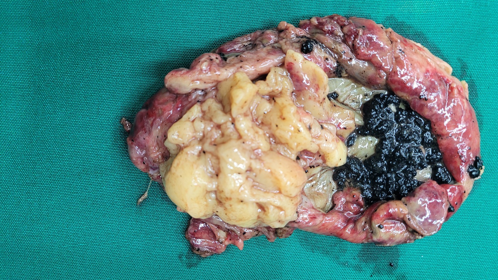
Finding the Lipoma: The ultrasound revealed a well-defined, homogeneous, echogenic lesion within the gallbladder wall. The characteristic features of this lesion, coupled with its fat composition, suggested a lipoma. To confirm the diagnosis and rule out other potential conditions, further imaging with a CT scan was conducted. The CT scan confirmed the presence of a lipoma, showing a fat-density lesion consistent with a benign tumor.
4)
Clinical Implications: While gallbladder lipomas are benign and often require no treatment, their presence can be concerning for both patients and clinicians due to the potential for misdiagnosis. It's crucial to differentiate these from more serious conditions such as gallbladder cancer or adenomas. In this case, the lipoma was confirmed to be benign, and the patient was advised on routine follow-up to monitor any changes.
Management and Follow-Up:
The management of gallbladder lipomas typically involves regular surveillance rather than immediate intervention. In this case, the patient was advised to undergo periodic imaging to ensure that the lipoma remained stable and did not show any signs of malignancy. Follow-up care is essential to confirm that the lipoma remains asymptomatic and does not grow, which could potentially cause issues.
Conclusion:
The discovery of a gallbladder lipoma, while rare, highlights the importance of thorough diagnostic evaluation in patients with gallbladder abnormalities. As this case illustrates, a careful approach involving various imaging modalities can help in accurate diagnosis and appropriate management.
For clinicians, it’s vital to be aware of such rare conditions and consider them in differential diagnoses when encountering atypical findings. For patients, understanding the nature of their condition can alleviate concerns and guide them towards the appropriate course of action.
We hope this case study provides valuable insights into the management of gallbladder lipomas and underscores the importance of vigilance in the diagnostic process. If you have any questions or would like to discuss this case further, feel free to reach out to us.
Stay tuned to our blog for more updates and case studies on various medical conditions and advancements in healthcare.